

 Hans Fried1, Roland Nitschke2, Kees van der Oord3, Maria M. Azevedo4,5
1. CRFS – Light Microscope Facility, German Center for Neurodegenerative Diseases (DZNE), Bonn, Germany
2. Life Imaging Center and Signalling Research Centre CIBSS, University of Freiburg, Germany
3. Nikon Europe BV, Amstelveen, Netherlands
4. Advanced Light Microscopy Scientific Platform, i3S – Instituto de Investigação e Inovação em Saúde | Porto, Portugal
5. University of Porto
Accurate multi‑colour imaging requires that signals from the same physical location co‑localise across channels. Chromatic aberration is a major source of spatial error in multi‑colour fluorescence microscopy. Therefore, rigorous co‑registration is essential for reliable interpretation of spatial relationships in biological specimens.
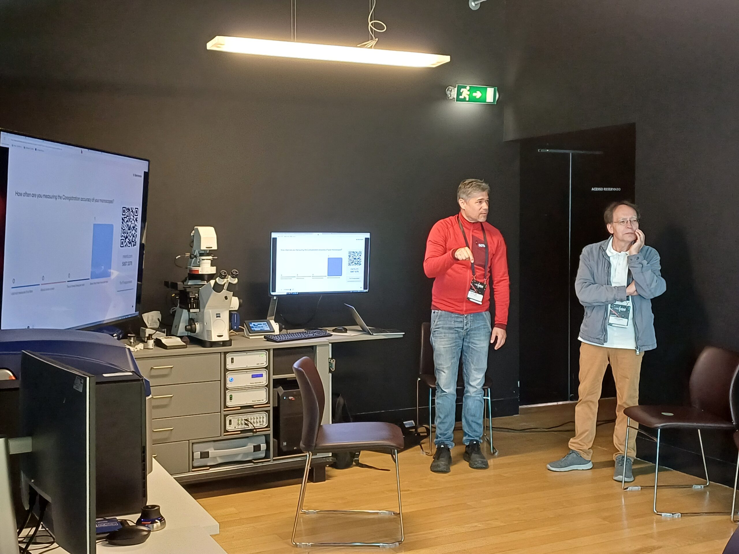
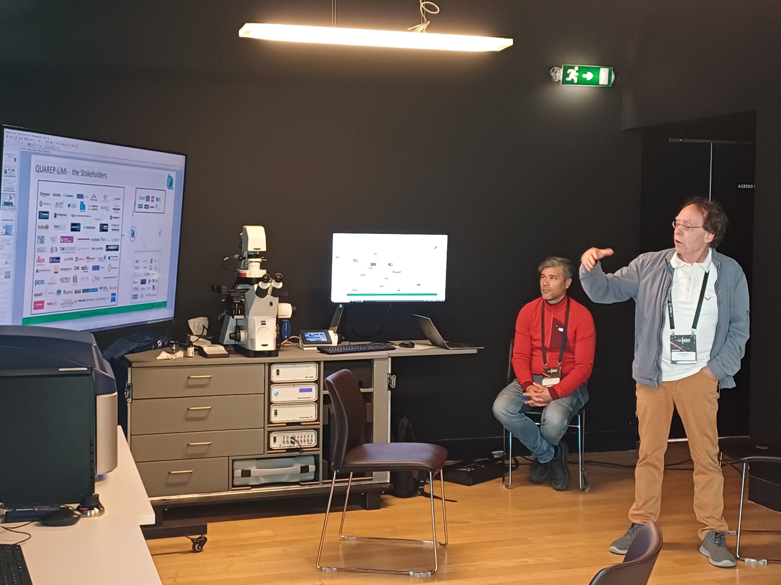
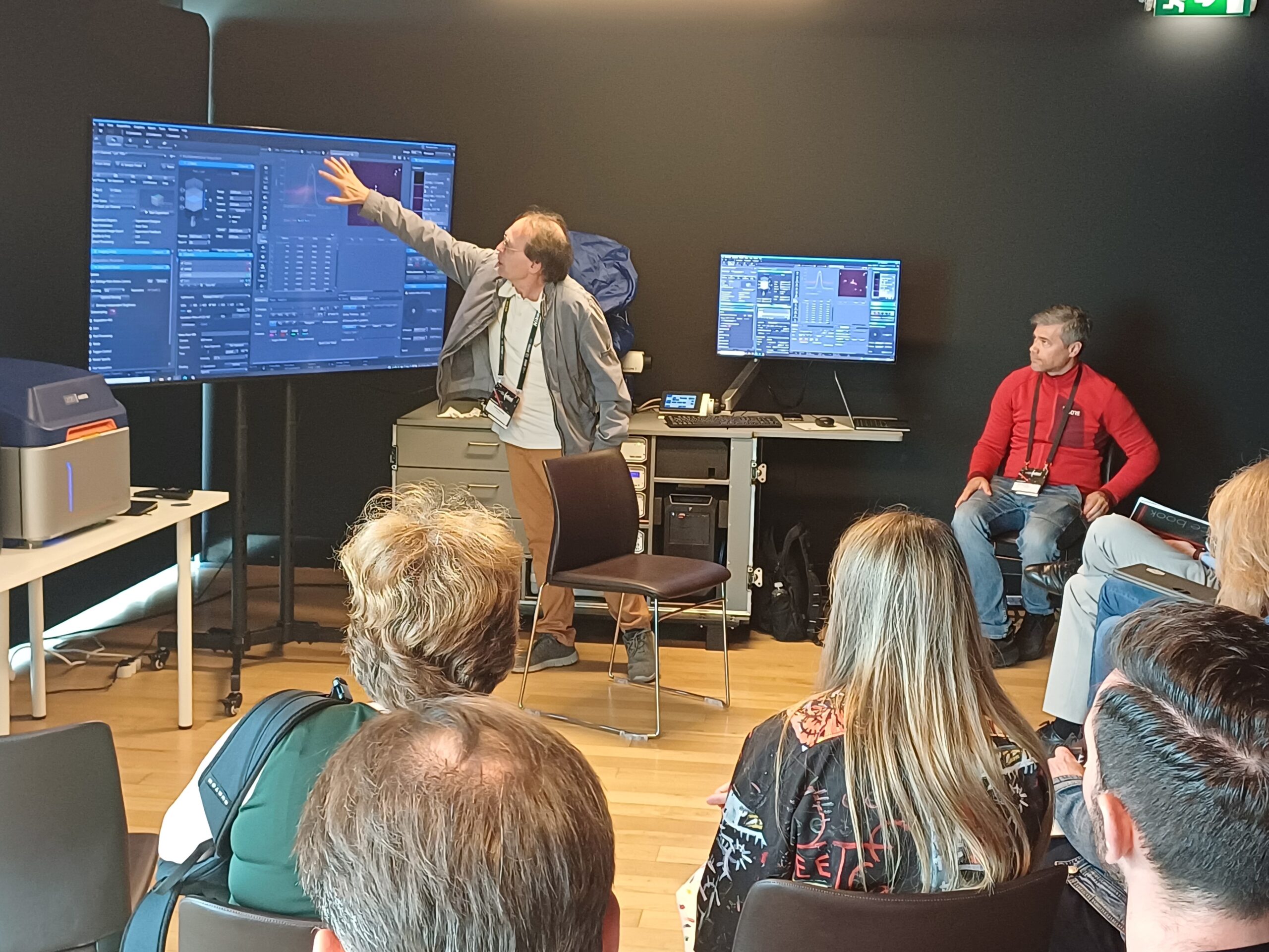
In this workshop, we will briefly explain the key steps of preparing a bead sample for co-registration assessment, show the main considerations for image acquisition (1), and show you how to analyze the obtained images using Fiji/ImageJ (2). We will also demonstrate how specific objective‑related factors can influence co‑registration performance. Finally, we will share practical hints and tips to support routine co‑registration analysis.
(1) Dauphin A., Azevedo, M., et al. Protocols.io, Ensuring accurate co-registration measurement for quality control of Single Point Confocal Laser Scanning Microscopes – V1, dx.doi.org/10.17504/protocols.io.q26g7yrj8gwz/v1
(2) Faklaris, O. et al. Quality assessment in light microscopy for routine use through simple tools and robust metrics. J Cell Biol 221, doi:10.1083/jcb.202107093 (2022).