
 Glyn Nelson, Ioannis Alexopoulos, Yury Belyaev
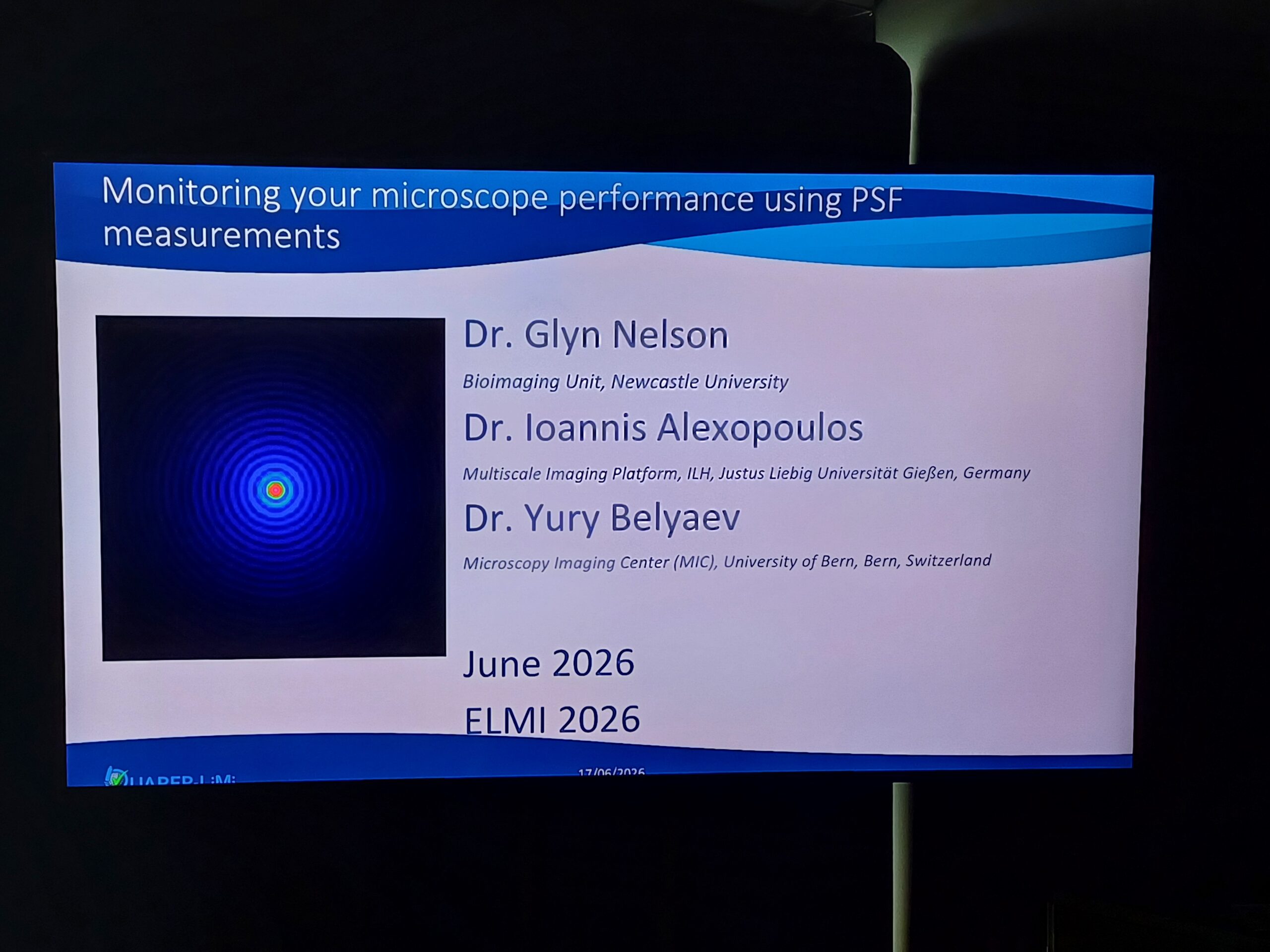
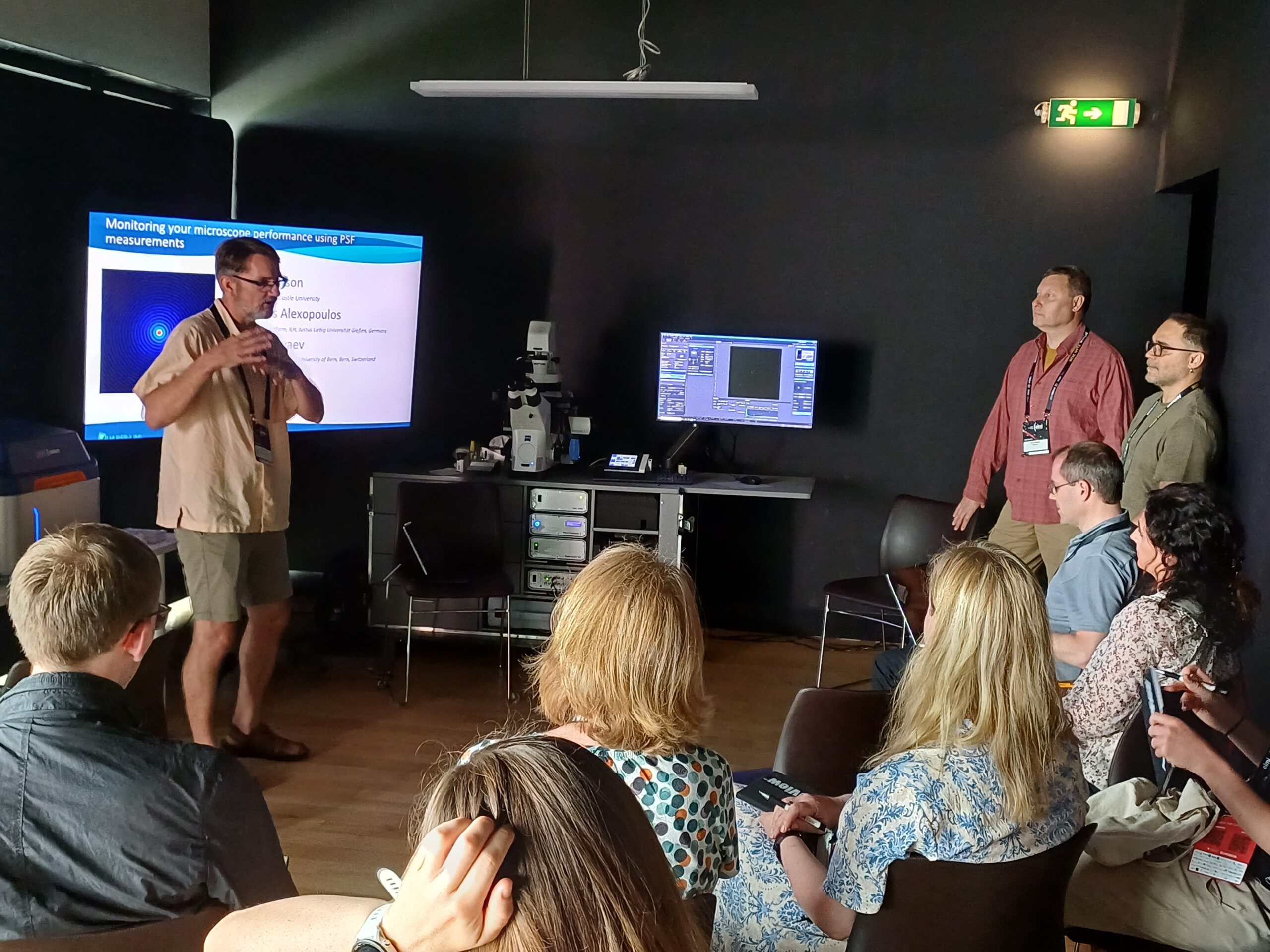
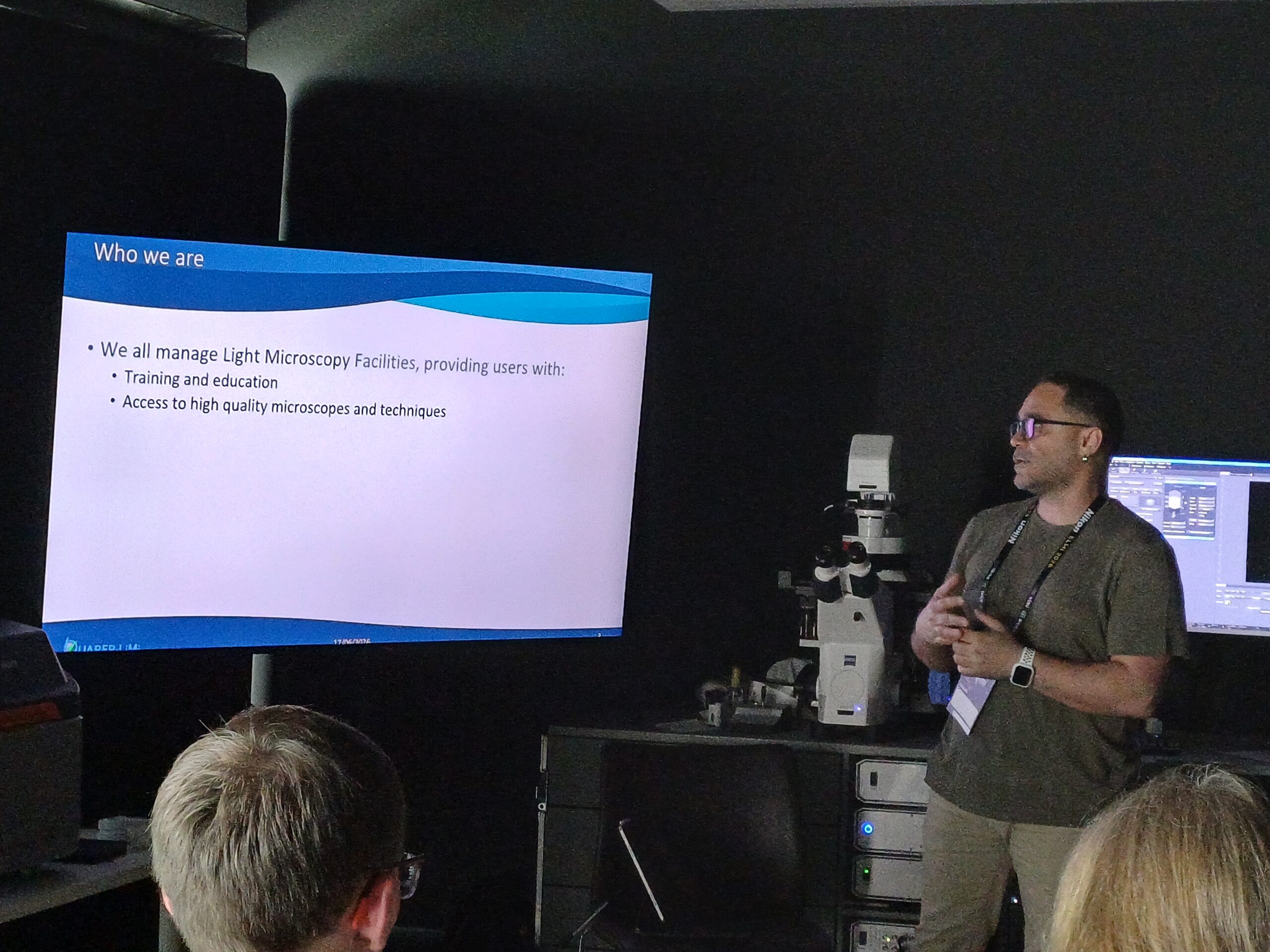
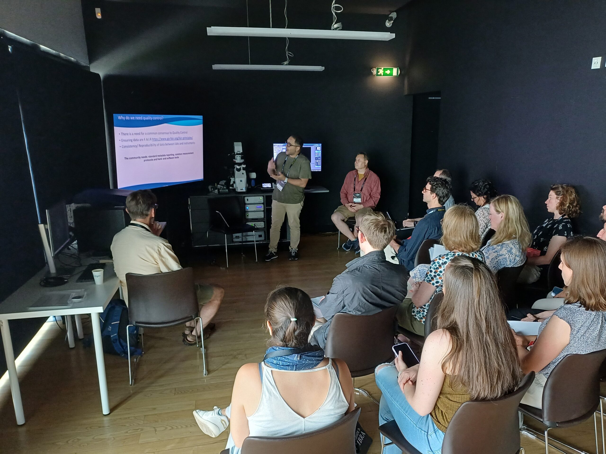
The Point Spread Function (PSF), which describes the response of an optical system to a point source, is a key quality control of a microscope. Regular measurements, conducted with consistent tools, methods, and protocols, along with the calculation of robust metrics enable the monitoring of the microscope performance and therefore ensure better reproducibility of scientific experiments. During this workshop, we will show briefly how to prepare a bead slide, how to perform an acquisition, and finally how to analyze the PSF with open-source tools (1, 2). We will give some tips to troubleshooting PSFs that have an abnormal shape or are far from the theoretical expected size. We will also demonstrate the upload of the analysis results to OMERO, a database that will help monitor PSFs over time. We will use the protocols and metrics that are defined in the framework of the QUAREP-LiMi consortium’s WG5 (3).
1. Faklaris O., et al. “Quality Assessment in Light Microscopy for Routine Use through Simple Tools and Robust Metrics.” Journal of Cell Biology 221, no. 11 (2022): e202107093. https://doi.org/10.1083/jcb.202107093.
2. https://github.com/MontpellierRessourcesImagerie/MetroloJ_QC
3. Nelson G., et al. Protocolos.io, Monitoring the point spread function for quality control of confocal microscopes , dx.doi.org/10.17504/protocols.io.bp2l61ww1vqe/v1
Britta Schroth-Diez, Kees van der Oorda Nikon Europe B.V.
b Light Microscopy Facility, Max Planck Institute of Molecular Cell Biology and Genetics, Dresden Germany
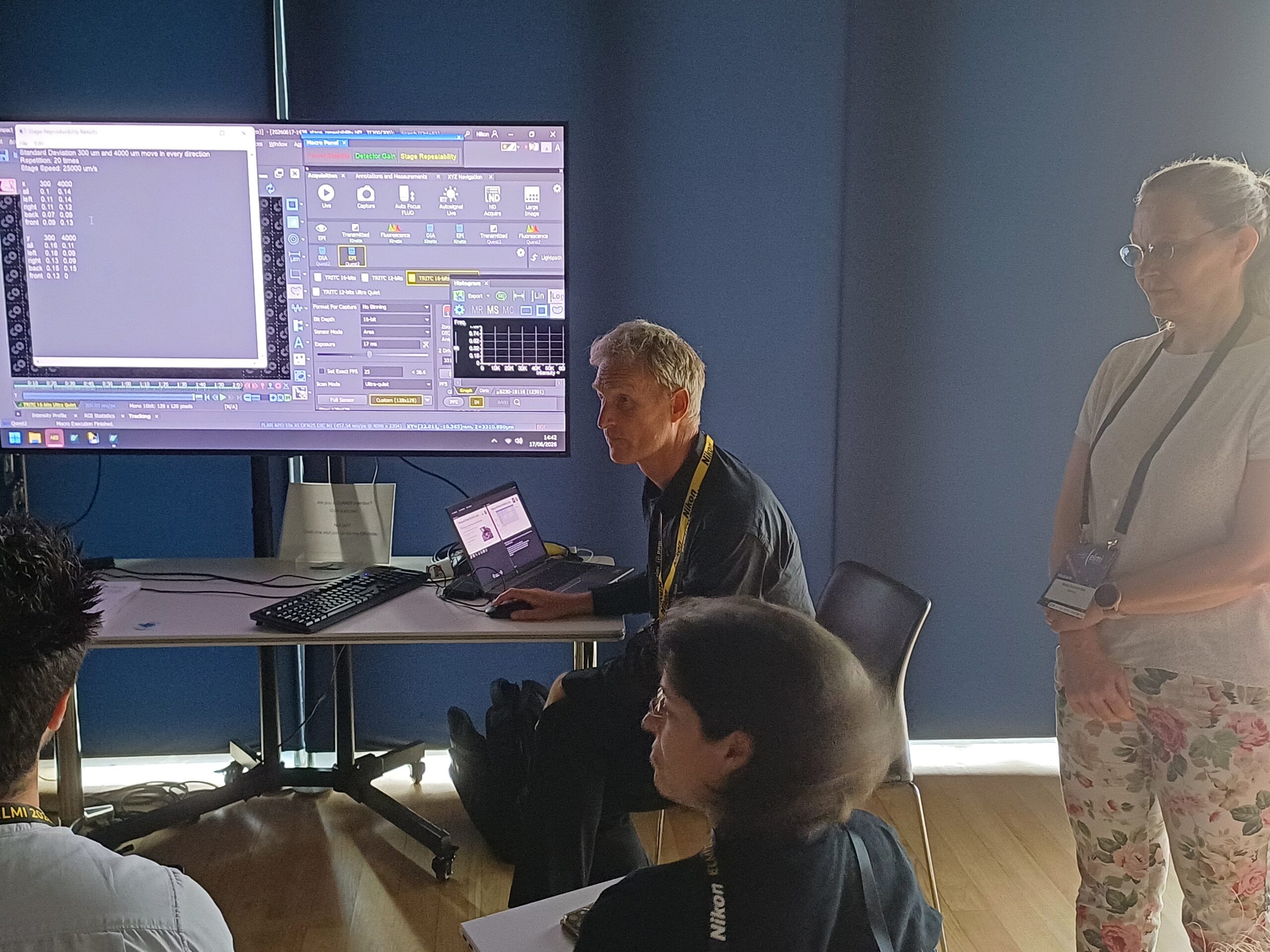
The QUAREP-LiMi Tool Kit [1] was developed by the QUAREP-LiMi community to reduce the time needed to run periodic quality assessment protocols and organize the performance data acquired. During this workshop we will present its basic features and latest developments.
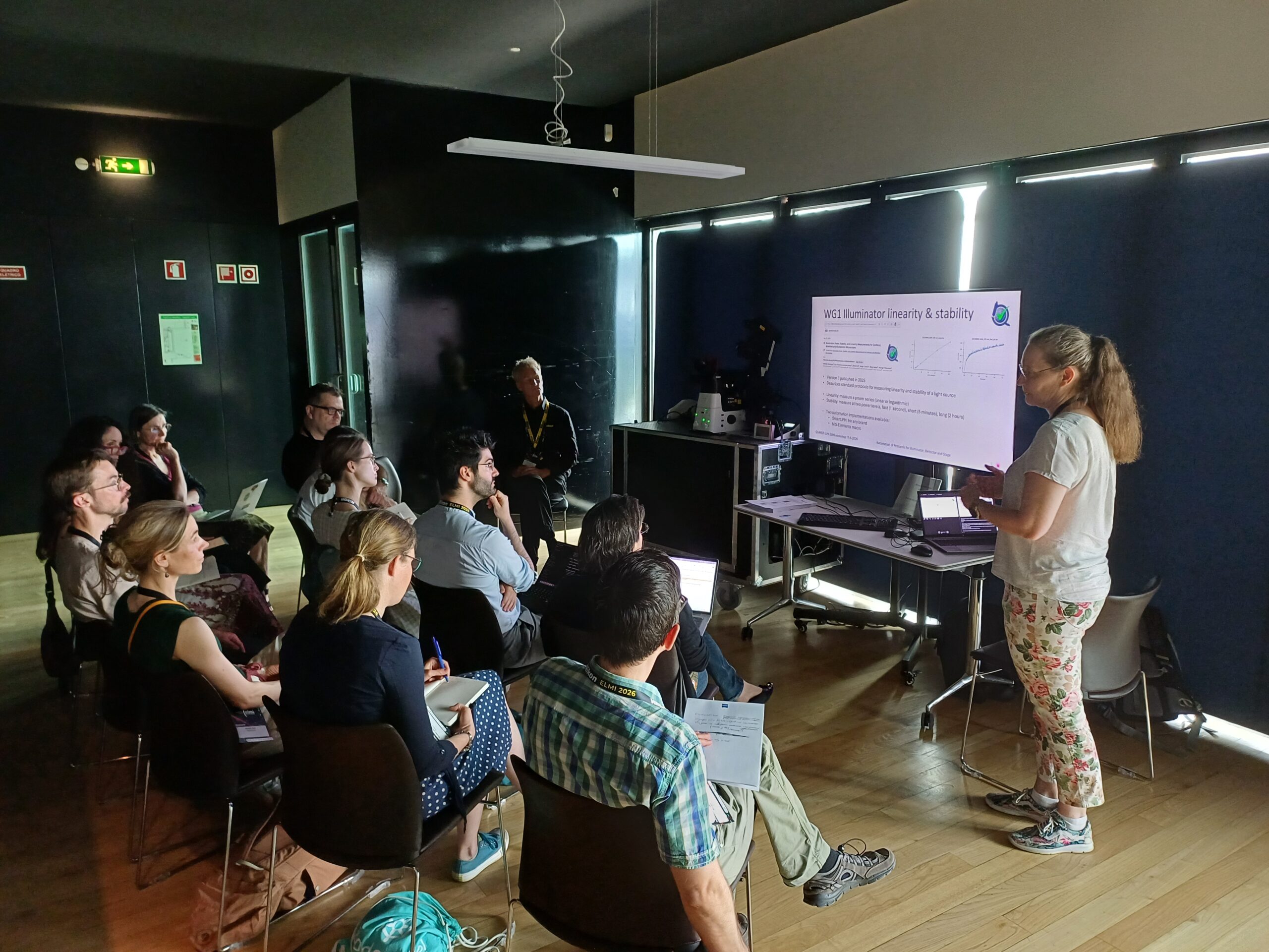
In accordance to the protocol defined by the QUAREP-LiMi WG 1 [2] the Tool Kit integrates user friendly interfaces for the assessment of illumination power and stability. These interfaces are already available for the Nikon and Zeiss acquistion software.
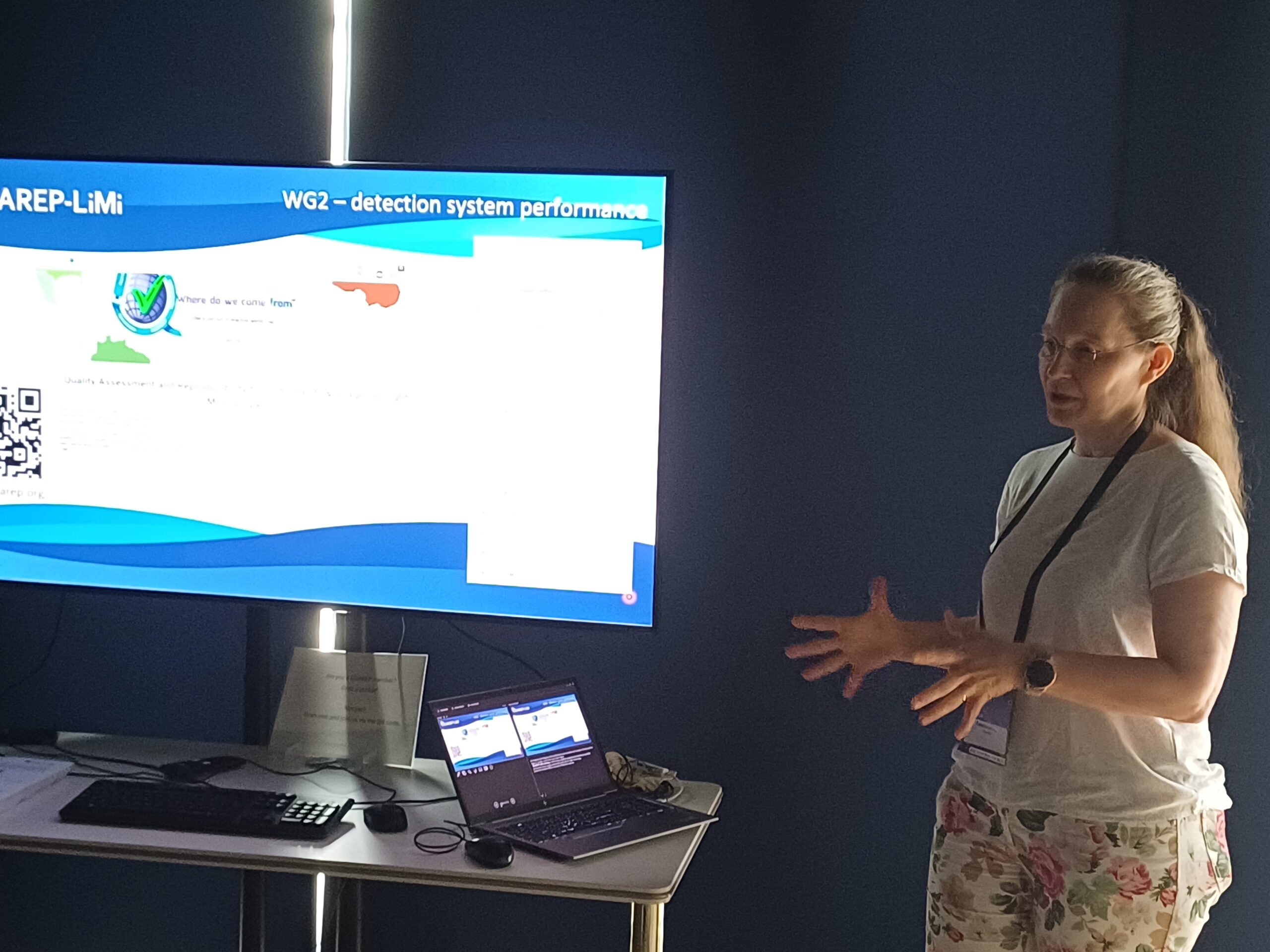
In addition, the Tool Kit includes support for the protocols for detector gain, dynamic range and noise assessment developed by the QUAREP-LiMi WG2 [2].
A highlight of the Tool Kit is its data browser. Thanks to the organized presentation of the performance metrics it is easier to diagnose problems – e.g. a small but steady power decrease over months– and take corrective and preventive actions before they become detrimental.
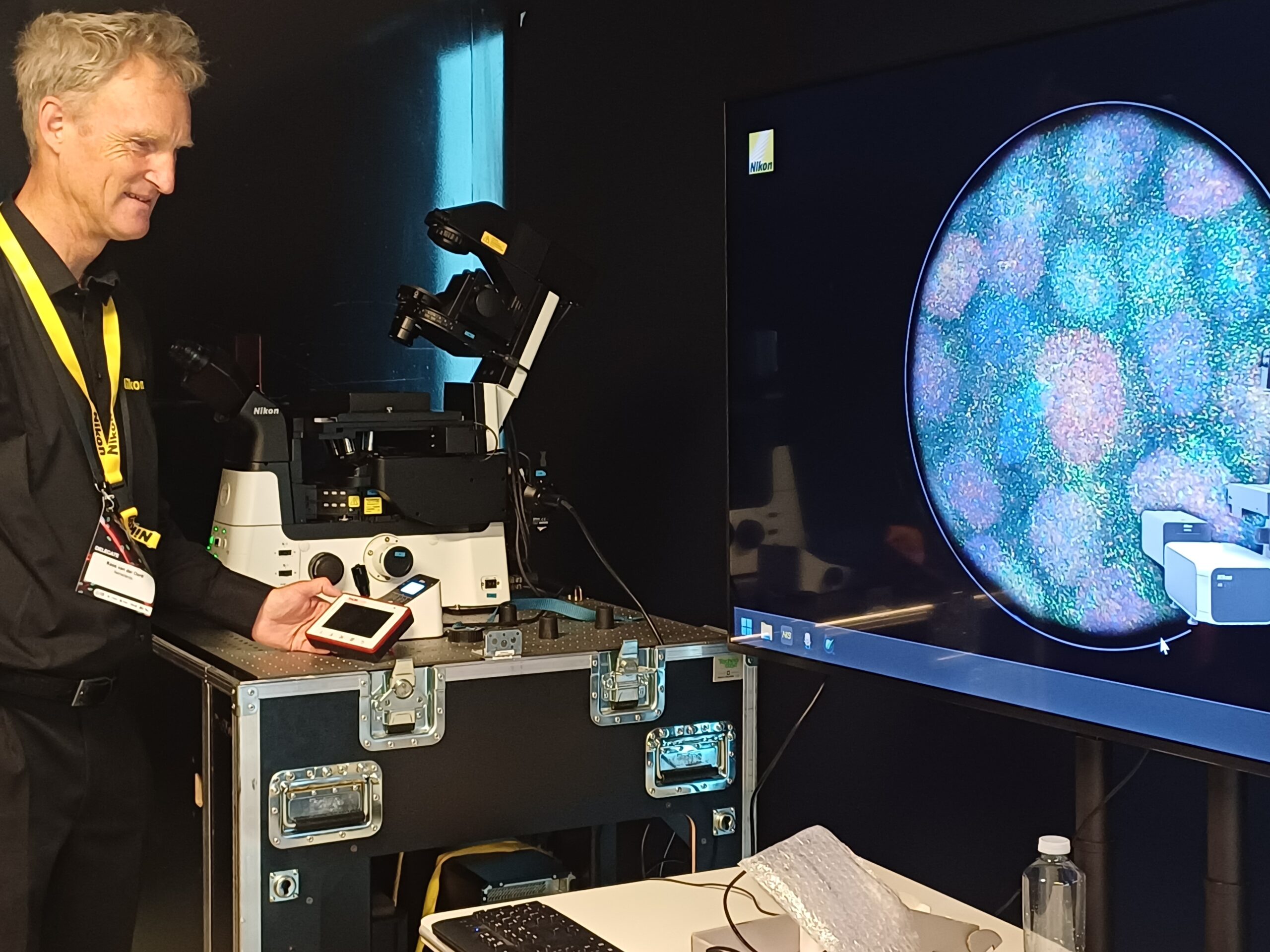
The Tool Kit functionality will be demonstrated during the workshop, where we will emphasize important details of the protocols.
[1] https://github.com/QUAREP-LiMi/QUAREP-LiMi-Tool-Kit
[2] https://www.protocols.io/workspaces/quarep-limi/publications
Britta Schroth-Diez, Kees van der Oord, Ruediger Bader1. Light Microscopy Facility, Max Planck Institute of Molecular Cell Biology and Genetics, Dresden Germany
2. Nikon Europe BV
3. Hamamatsu Photonics Europe
QUAREP-LiMi Working Group 2 (WG2) developed a set of protocols [1] which focuses on the characterization and monitoring of the performance of light microscope detection systems, which collects light from the sample at the microscope and helps to convert the “arbitrary digital units” (ADU) provided by the microscope into “number of photons” detected.
The protocols collection is built around the photon transfer curve (PTC) method [2]. The goals of these protocols are to achieve 1) experiment quality control, 2) monitoring instrument quality over time, and 3) detailed detection system characterization. The protocols include both point and area detectors, allowing researchers to select appropriate methods based on their specific detection systems.
During the workshop, we will demonstrate how to use the protocols to acquire images and upload them in the software [3] to characterize the photon conversation factor (PCF photons/ADU), readnoise, background and dynamic range of a light microscopy detection system. The set includes protocols on sample preparation, data acquisition, and analysis. Furthermore, we will demonstrate its integration in the QUAREP-LiMi Tool Kit [4].
[1] Characterization of the Photon Conversion Factor, Noise, and Dynamic Range of Light Microscope Detection Systems – https://dx.doi.org/10.17504/protocols.io.14egn61pyl5d/v1
[2] Janesick JR. 2007. Photon Transfer. SPIE. https://dx.doi.org/10.1117/3.725073
[3] McFadden D. 2022. GUI Calibration Tool. https://github.com/bionanoimaging/NanoImagingPack/releases
[4] QUAREP-LiMi Tool Kit, https://github.com/QUAREP-LiMi/QUAREP-LiMi-Tool-Kit

QUAREP-LiMi WG3 was pleased to host a webinar presented by Daniel Schröder on the topic of the "Universal Laser Engine" with an improved homogeneous illumination profile.
The webinar introduced a versatile laser excitation platform designed for advanced fluorescence microscopy and highlighted its applications across multiple imaging modalities.
Abstract
The Universal Laser Engine is a user-friendly laserbox designed to provide a versatile excitation for advanced fluorescence microscopy. In a single box, it holds up to 6 laser wavelengths operating in modes ranging from picosecond-pulsed to continuous-wave, enabling its use across a wide range of imaging modalities. The Universal Laser Engine has been successfully tested in widefield microscopy, super-resolution localization microscopy, total internal reflection fluorescence (TIRF), and fluorescence lifetime imaging microscopy (FLIM). In addition, the Universal Laser Engine incorporates a novel speckle-suppression technology that generates a highly homogeneous flat-top hat profile that is applicable for high-speed imaging at exposure times down to microseconds. The system provides various trigger inputs and two fiber outputs for seamless integration into both existing and new microscopes. By combining flexibility, compact design, and state-of-the-art illumination uniformity, the Universal Laser Engine offers a powerful and adaptable solution for simplifying and modernizing fluorescence microscopy instrumentation.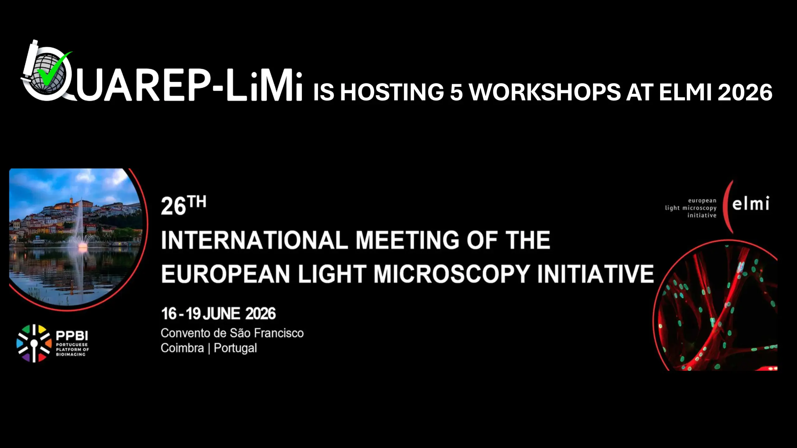
We are pleased to announce that the QUAREP-LiMi consortium will be presenting five specialized workshops at the upcoming European Light Microscopy Institute (ELMI) meeting. Our sessions are designed to provide researchers with frameworks for improving image quality, ensuring data reproducibility, and implementing rigorous quality control standards in microscopy. The workshops will cover five critical pillars of instrument characterization:
-
The QUAREP-LiMi Tool Kit (WGs 1 & 2):
Discover how to streamline periodic quality assessments. We will present the latest developments in the Tool Kit, including user-friendly software tools for illumination power and stability (compatible with Nikon and Zeiss), as well as its data browser for long-term performance monitoring. -
Detector Performance & Photon Characterization (WG2):
Learn how to transition from "arbitrary digital units" (ADU) to actual photon counts using the Photon Transfer Curve (PTC) method. We will demonstrate protocols for characterizing photon conversion factors, read noise, and dynamic range for both point and area detectors. -
Assessing the co-registration accuracy of a microscope (WG4):
Chromatic aberration is a major source of spatial error in multi‑colour fluorescence microscopy. This workshop will show practical hints and tips to perform routine co‑registration measurement and analysis. -
Point Spread Function (PSF) Analysis (WG5):
This workshop provides a practical guide to PSF measurement—from beads slide preparation and image acquisition to analysis using open-source tools and integration with OMERO for longitudinal monitoring. -
The Light-Microscopy Metadata Model (LiMi-Model, WG7):
In collaboration with BioImaging North America, 4D Nucleome, and OME, we will introduce the LiMi-Model. This framework aims to harmonize the description of hardware and acquisition settings to ensure datasets are FAIR (Findable, Accessible, Interoperable, and Reusable) by design.

We warmly welcome our first member from Nigeria, Dr Olubunmi Ayobami Balogun, from the University of Ibadan who just joined Working Group 1, 4 and 12.
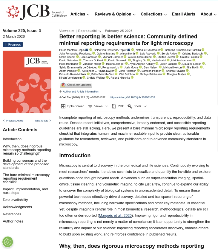
A new article, under the guidance of QUAREP-LiMi WG 11, on the minimal reporting requirements for light microscopy data has been published in the Journal of Cell Biology. You find the article here.
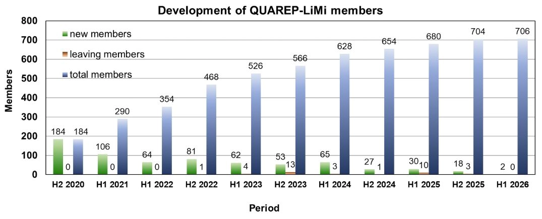
QUAREP-LiMi has reached another milestone. By the end of 2025 more than 700 members have joined QUAREP-LiMi. When we launched this initiative, we never imagined the community would grow to include so many members from 46 different countries around the globe. A heartfelt thank‑you to all our members for your continued support and contributions.
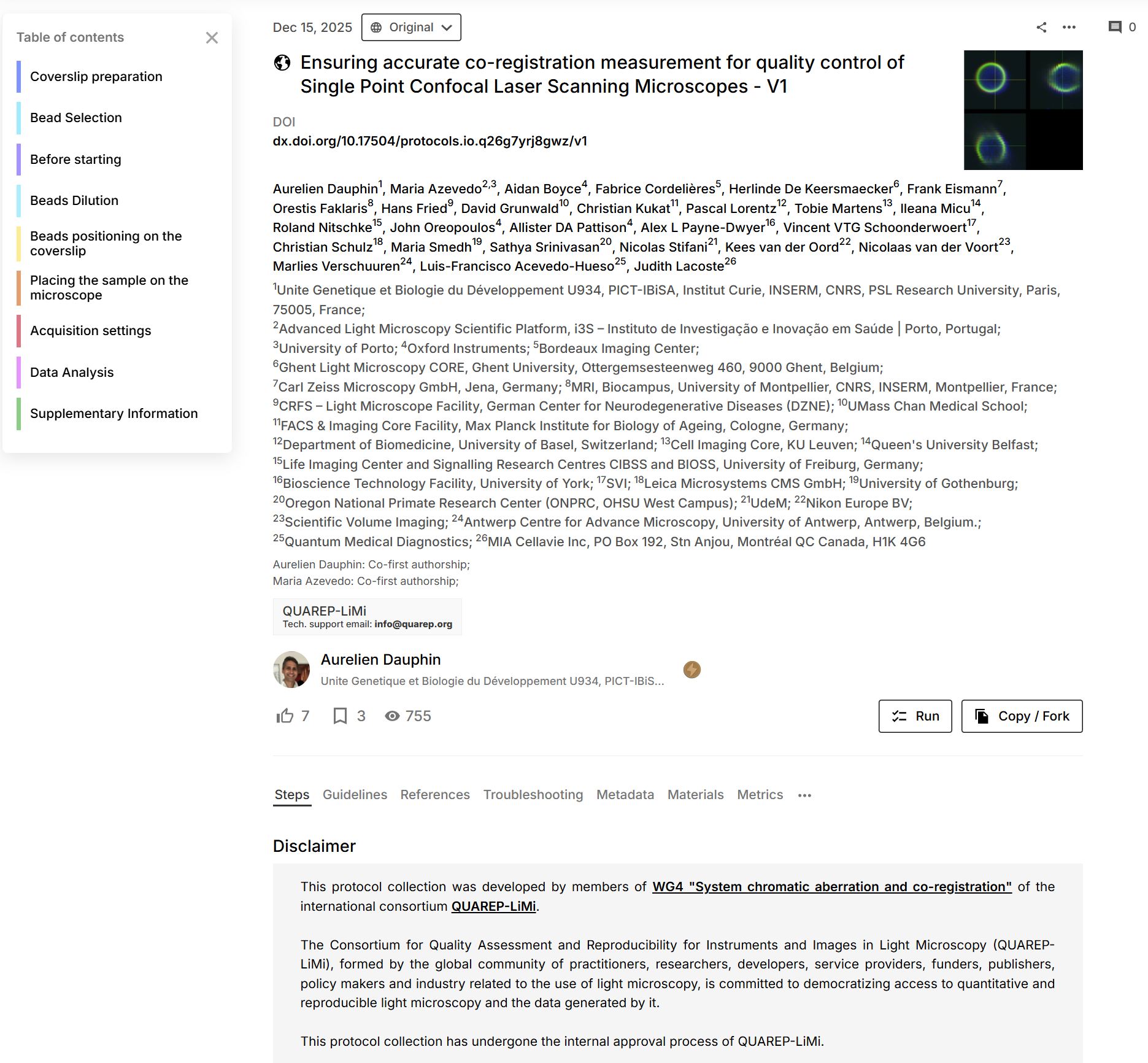
We are thrilled that WG 4 - System chromatic aberration and co-registration Power published its first protocol "Ensuring accurate co-registration measurement for quality control of Single Point Confocal Laser Scanning Microscopes - V1". The protocol is featured on protocols.io

The project “Increased Resource Efficiency in Light Microscopy by Quality Control and Standards” has been selected for funding by the Deutsche Forschungsgemeinschaft (DFG, German Research Foundation). The project supports a collaborative effort led by Dr Roland Nitschke (University of Freiburg), Dr Hella Hartmann (TU Dresden), and Dr Janina Hanne (GerBI-GMB, Konstanz). They will receive approximately one million euros in joint funding for 3 positions over 3 years as part of a nationwide initiative to improve the sustainability of large-scale scientific equipment use.
Three positions are available in this project from now on. For more information download the PDF file. If you want to apply for one of these position use our application form.

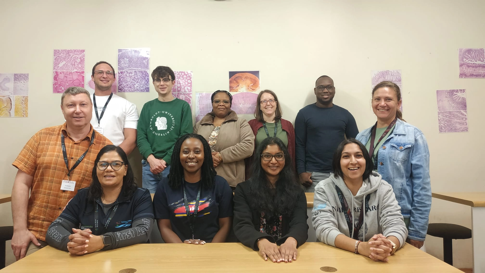
In April 2025, Yury Belyaev from the Microscopy Imaging Center (MIC) at the University of Bern, and a member of QUAREP-LiMi Working Group 5 (WG5), visited the Central Analytical Facilities (CAF) Microscopy Unit at Stellenbosch University, South Africa, as part of the Global BioImaging (GBI) International Job Shadowing Program
During his visit, Yury Belyaev collaborated with Lize Engelbrecht, manager of the CAF Microscopy Unit, to organize a hands-on microscopy workshop. The training covered wide-field and confocal microscopy, image analysis, and microscope maintenance. The event was generously supported by the CoRE Genomics for Health in Africa Scientific Exchange Program and Chroma
The workshop brought together participants from across South Africa and provided an excellent platform for Yury to showcase QUAREP-LiMi's activities, along with other international microscopy and image processing initiatives. A dedicated maintenance day also allowed for the presentation of protocols developed by QUAREP-LiMi, particularly those from WG5 in which Yury is actively involved.