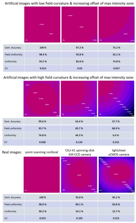
New QUAREP-LiMi Protocol: Monitoring Field Homogeneity Working Group 3 (Field Homogeneity) is pleased to share a new open-access protocol: “Monitoring Field Homogeneity”, authored by colleagues from institutions across Europe, Canada, and beyond. Field homogeneity — the uniformity of illumination and detection across the field of view — is a cornerstone of quantitative fluorescence imaging. Changes […]
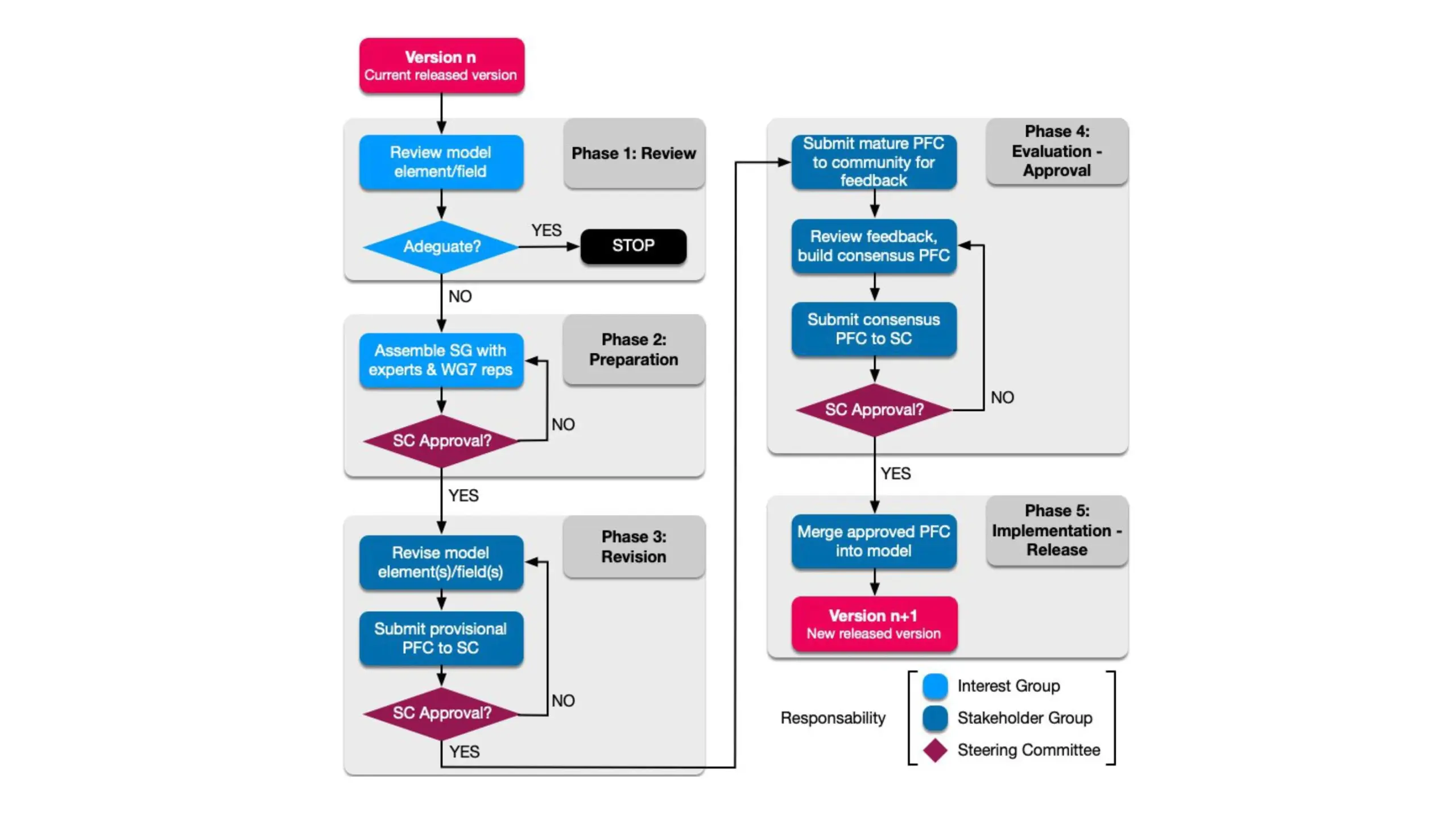
WG7-Metadata is proud to announce the publication of the LiMi-Model Revision Best Practices. This document will govern all future revisions of the LiMi-Model, the QUAREP-LiMi standard for describing microscopy hardware, acquisition settings, and quality-control metrics. As depicted in the figure, the Best Practices lay out a five-phase process — Review, Preparation, Revision, Evaluation and Approval, […]
We are proud to announce that QUAREP-LiMi WG7-Metadata has launched the LiMi-model Website, the new home for the Light Microscopy Model — a community-built, FAIR-compliant framework for describing microscopy hardware, acquisition settings, and quality-control metrics. The site covers the model’s purpose and deliverables, and shows how to get involved, whether by exploring the model, testing […]
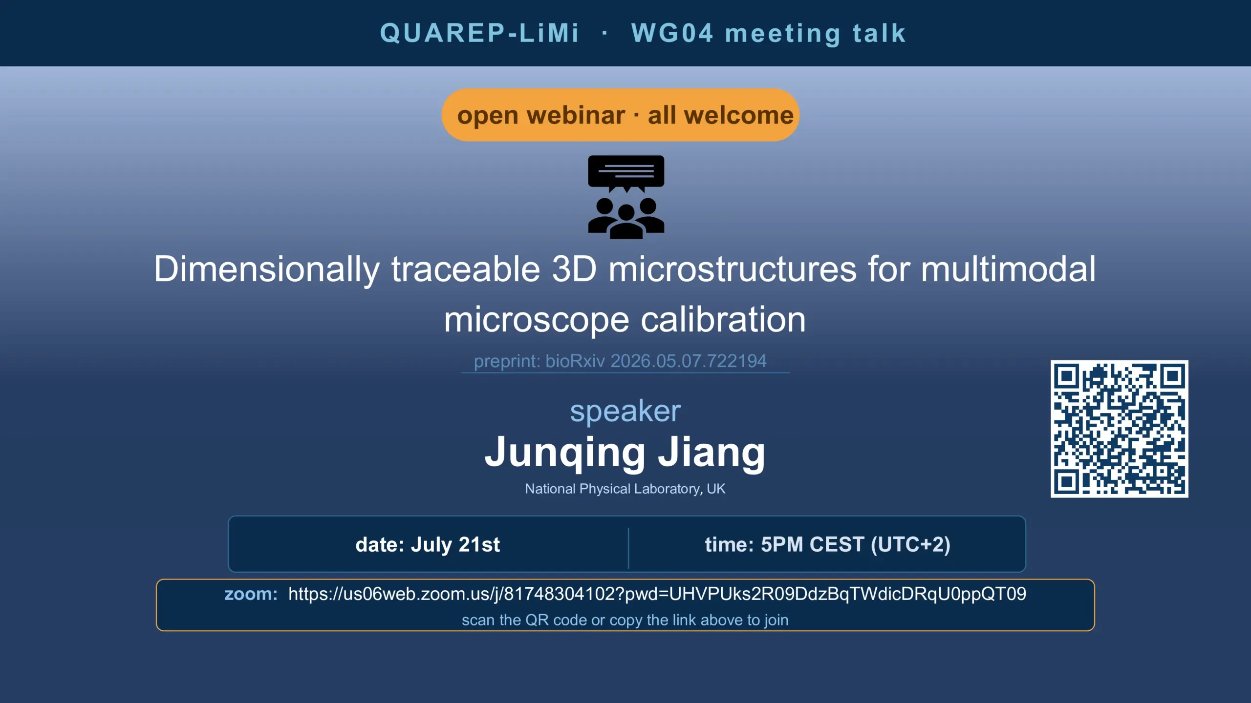
Open Webinar — QUAREP-LiMi WG4 Join us on July 21, 5PM CEST, as Junqing Jiang (NPL, UK) presents: “Dimensionally Traceable 3D Microstructures for Multimodal Microscope Calibration” Open to everyone working in microscopy, imaging, or metrology! Scan the QR code on the flyer or join via Zoom: https://us06web.zoom.us/j/81748304102?pwd=UHVPUks2R09DdzBqTWdicDRqU0ppQT09

Nataliia Aleksandrova Arnica Kaiping Jakub Pospíšil In exciting news from QUAREP-LiMi, three new staff members have joined forces as core QUAREP staff, for the first time in its history, to accelerate the project in various capacities. They were welcomed into the community with a vibrant meet and greet on 2. July. 2026. If you are […]
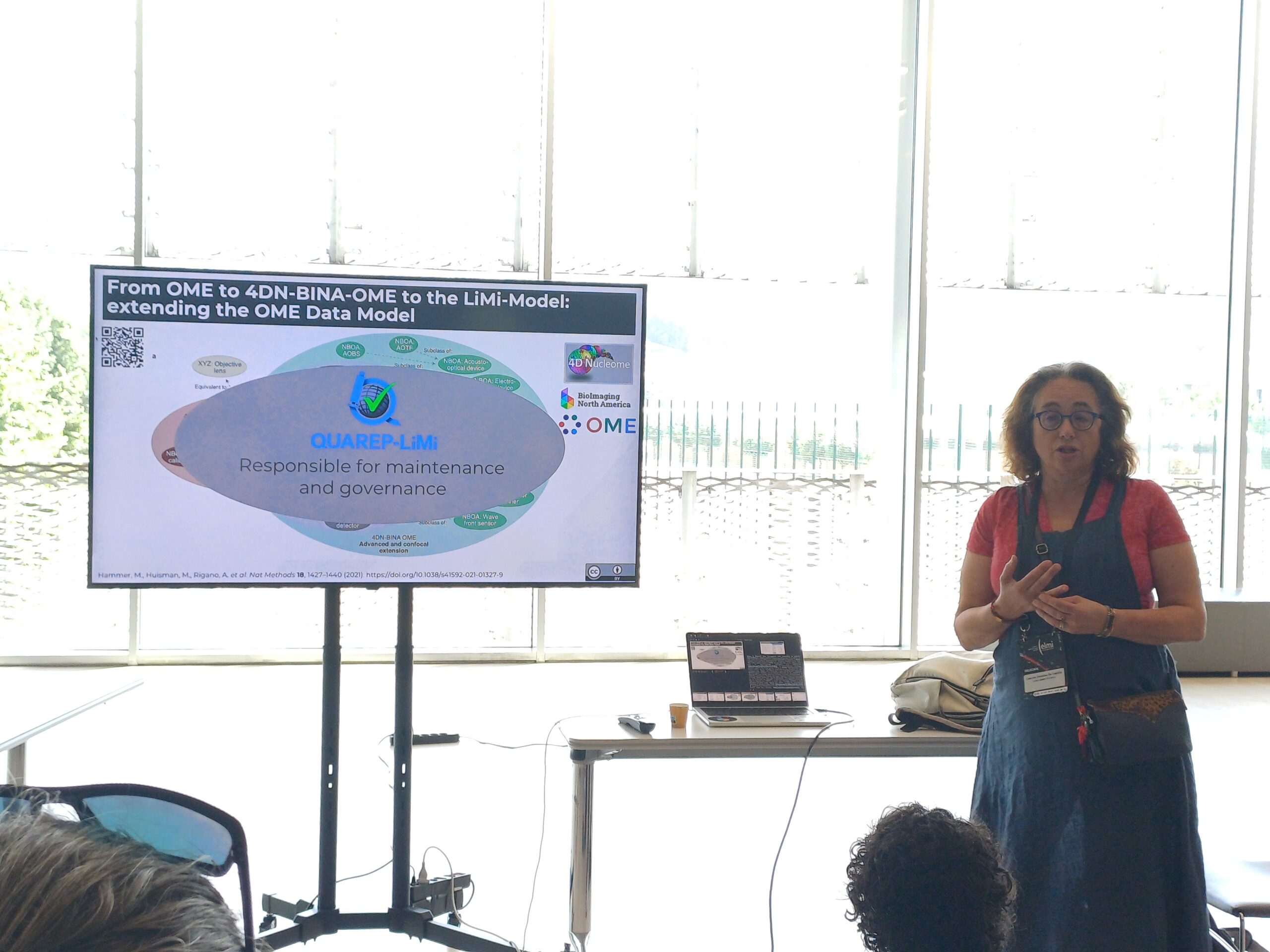
Caterina Strambio De Castillia Presenter(s): Caterina Strambio-De-Castillia *1 *- the workshop was presented on behalf of QUAREP-LiMi WG7-Metadata. 1. Program in Molecular Medicine, UMass Chan Medical School A major output of QUAREP-LiMi is the Light-Microscopy Metadata Model (LiMi-Model) [1, 2], developed by the QUAREP-LiMi Metadata Working Group (WG7) in collaboration with BioImaging North America, the […]
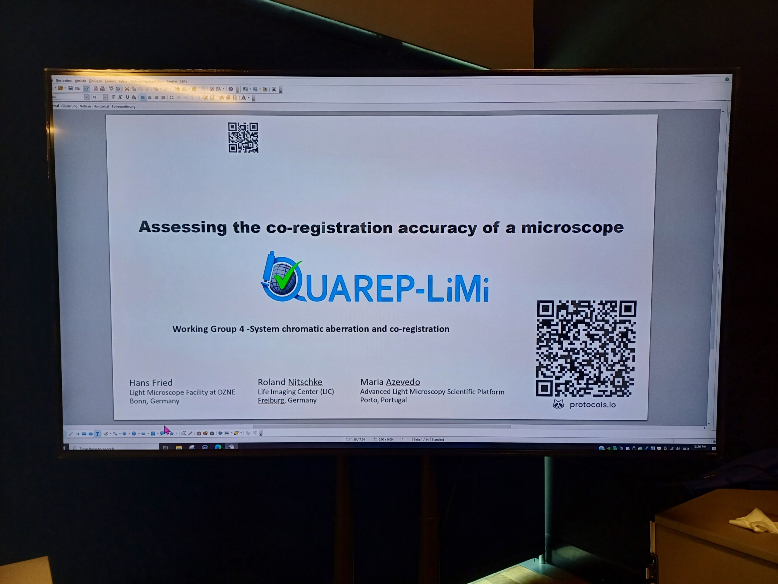
Hans Fried1, Roland Nitschke2, Kees van der Oord3, Maria M. Azevedo4,5 1. CRFS – Light Microscope Facility, German Center for Neurodegenerative Diseases (DZNE), Bonn, Germany 2. Life Imaging Center and Signalling Research Centre CIBSS, University of Freiburg, Germany 3. Nikon Europe BV, Amstelveen, Netherlands 4. Advanced Light Microscopy Scientific Platform, i3S – Instituto de Investigação […]
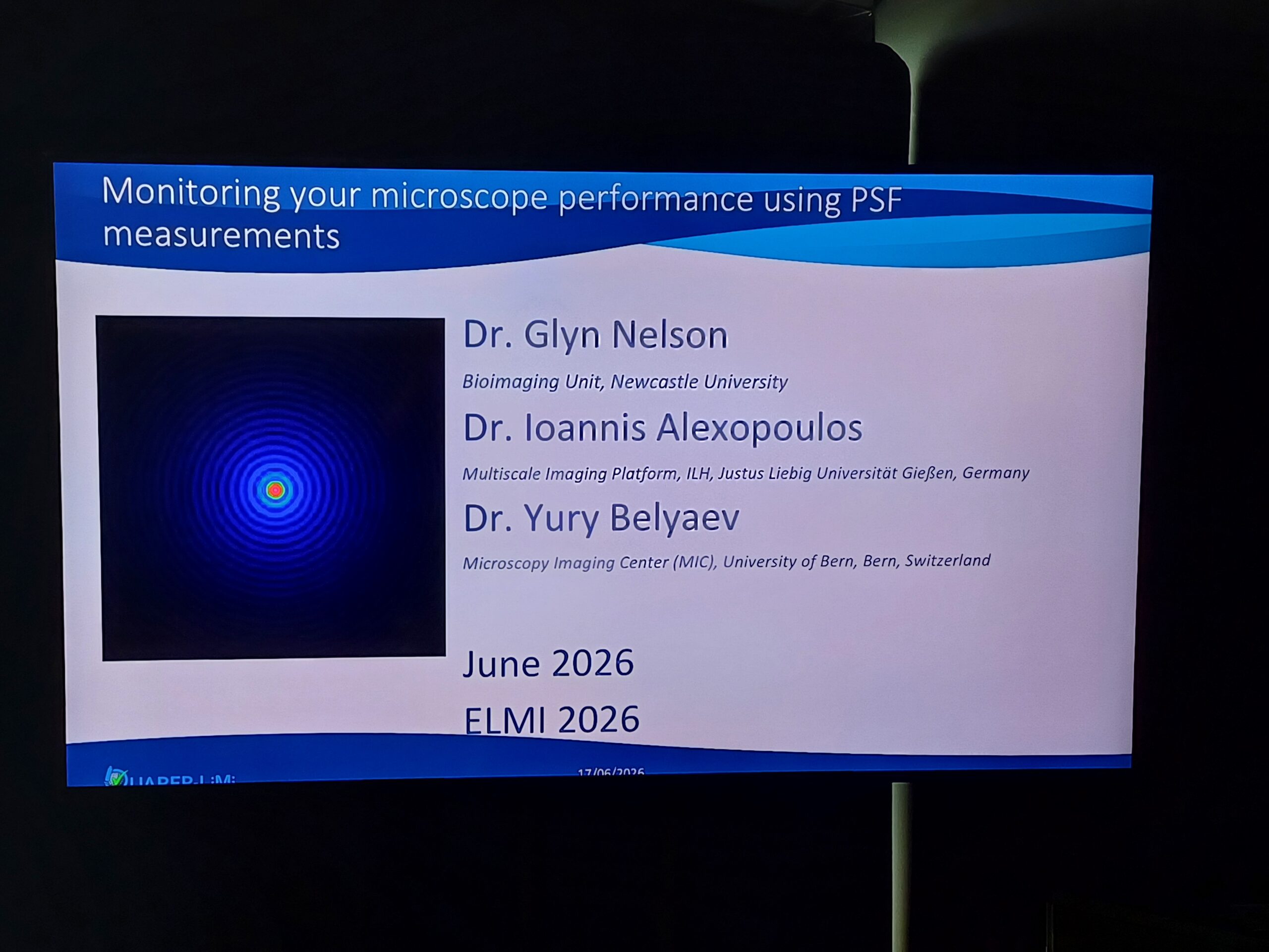
Glyn Nelson, Ioannis Alexopoulos, Yury Belyaev The Point Spread Function (PSF), which describes the response of an optical system to a point source, is a key quality control of a microscope. Regular measurements, conducted with consistent tools, methods, and protocols, along with the calculation of robust metrics enable the monitoring of the microscope performance and […]
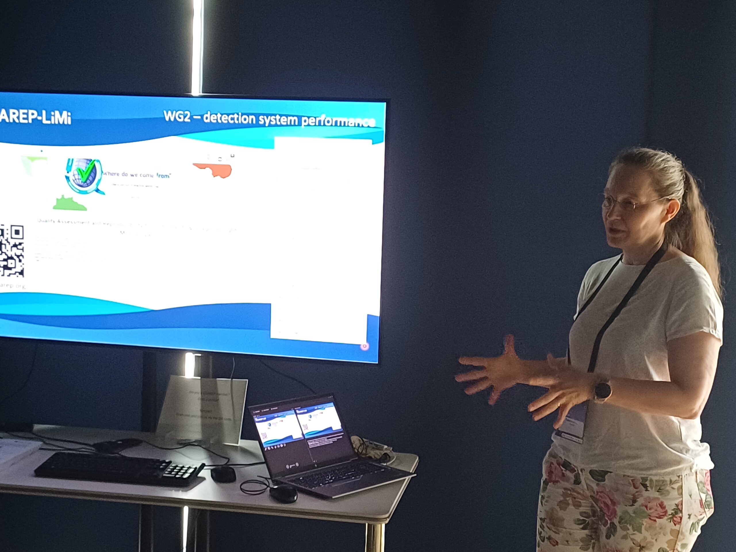
Britta Schroth-Diez, Kees van der Oord a Nikon Europe B.V. b Light Microscopy Facility, Max Planck Institute of Molecular Cell Biology and Genetics, Dresden Germany The QUAREP-LiMi Tool Kit [1] was developed by the QUAREP-LiMi community to reduce the time needed to run periodic quality assessment protocols and organize the performance data acquired. During […]
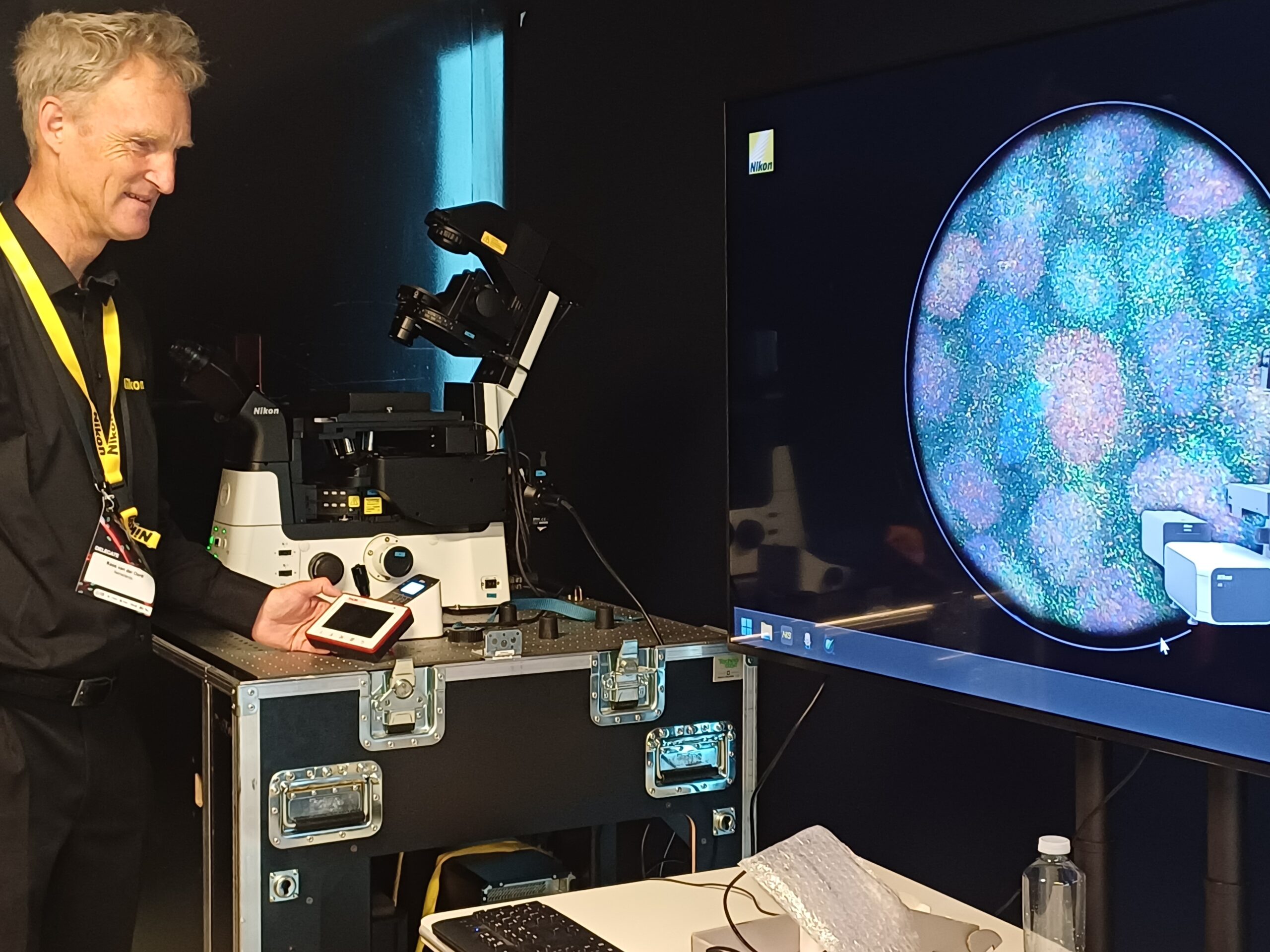
Britta Schroth-Diez, Kees van der Oord, Ruediger Bader 1. Light Microscopy Facility, Max Planck Institute of Molecular Cell Biology and Genetics, Dresden Germany 2. Nikon Europe BV 3. Hamamatsu Photonics Europe QUAREP-LiMi Working Group 2 (WG2) developed a set of protocols [1] which focuses on the characterization and monitoring of the performance of light […]

QUAREP-LiMi WG3 was pleased to host a webinar presented by Daniel Schröder on the topic of the “Universal Laser Engine” with an improved homogeneous illumination profile. The webinar introduced a versatile laser excitation platform designed for advanced fluorescence microscopy and highlighted its applications across multiple imaging modalities. Abstract The Universal Laser Engine is a user-friendly […]
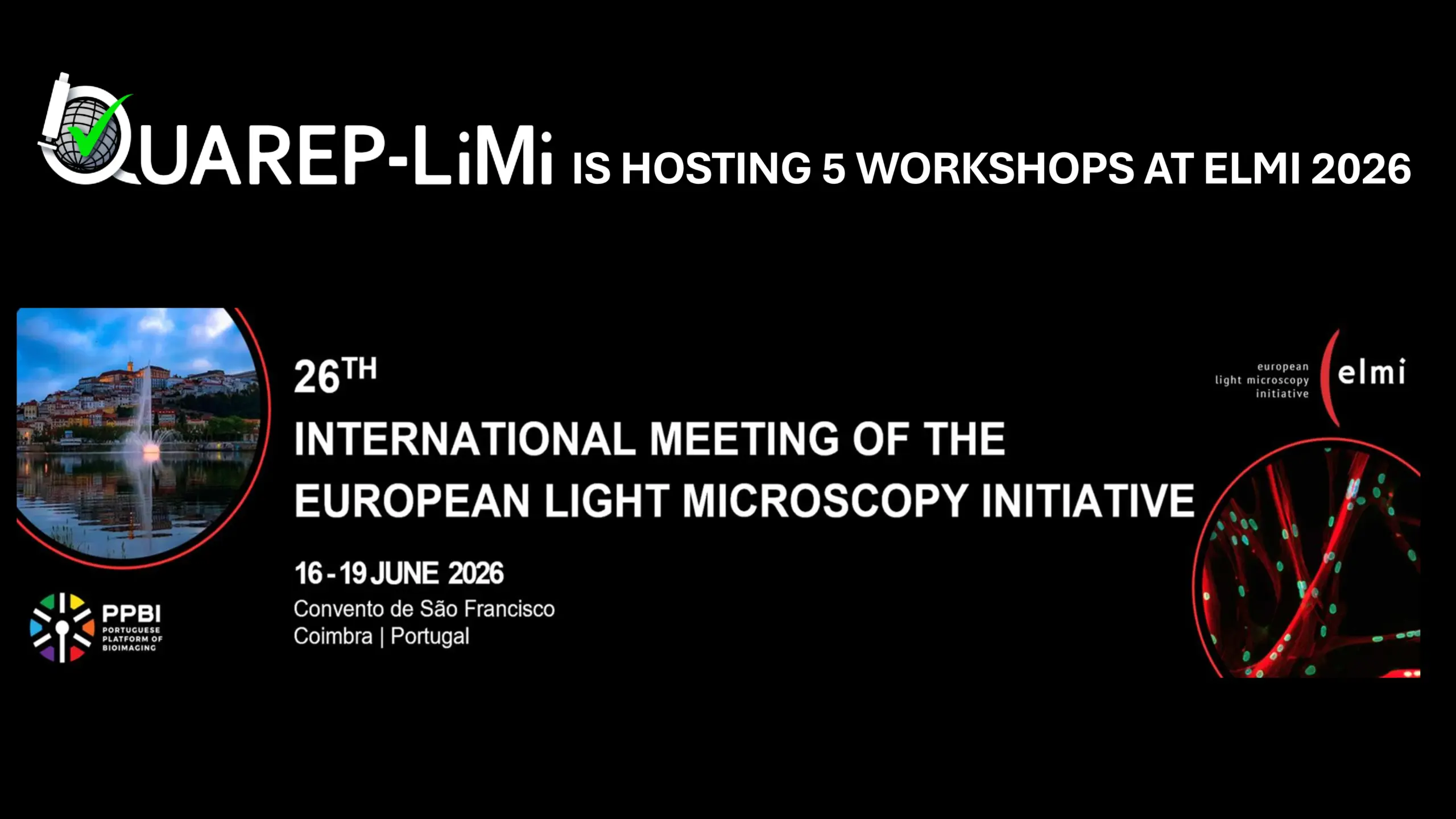
We are pleased to announce that the QUAREP-LiMi consortium will be presenting five specialized workshops at the upcoming European Light Microscopy Institute (ELMI) meeting. Our sessions are designed to provide researchers with frameworks for improving image quality, ensuring data reproducibility, and implementing rigorous quality control standards in microscopy. The workshops will cover five critical pillars […]

We warmly welcome our first member from Nigeria, Dr Olubunmi Ayobami Balogun, from the University of Ibadan who just joined Working Group 1, 4 and 12.
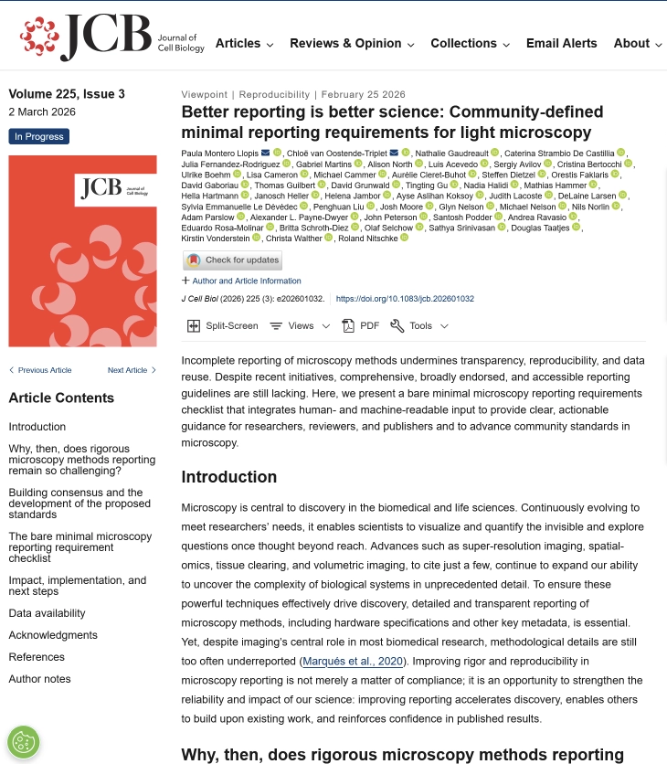
A new article, under the guidance of QUAREP-LiMi WG 11, on the minimal reporting requirements for light microscopy data has been published in the Journal of Cell Biology. You find the article here.
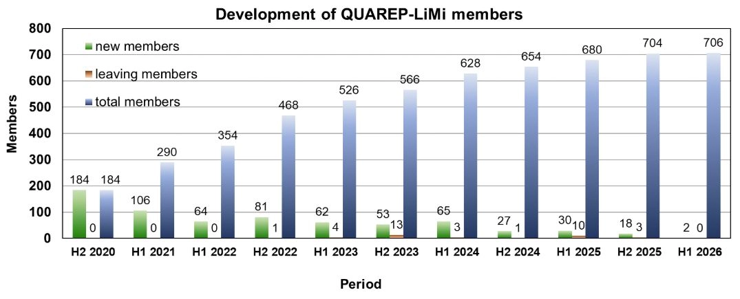
QUAREP-LiMi has reached another milestone. By the end of 2025 more than 700 members have joined QUAREP-LiMi. When we launched this initiative, we never imagined the community would grow to include so many members from 46 different countries around the globe. A heartfelt thank‑you to all our members for your continued support and contributions.
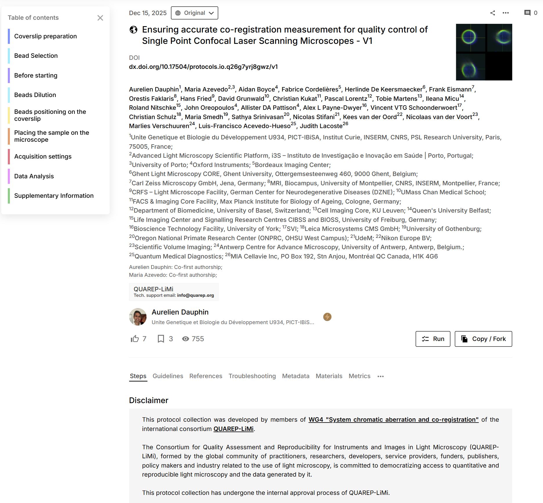
We are thrilled that WG 4 – System chromatic aberration and co-registration Power published its first protocol “Ensuring accurate co-registration measurement for quality control of Single Point Confocal Laser Scanning Microscopes – V1“. The protocol is featured on protocols.io

The project “Increased Resource Efficiency in Light Microscopy by Quality Control and Standards” has been selected for funding by the Deutsche Forschungsgemeinschaft (DFG, German Research Foundation). The project supports a collaborative effort led by Dr Roland Nitschke (University of Freiburg), Dr Hella Hartmann (TU Dresden), and Dr Janina Hanne (GerBI-GMB, Konstanz). They will receive approximately […]
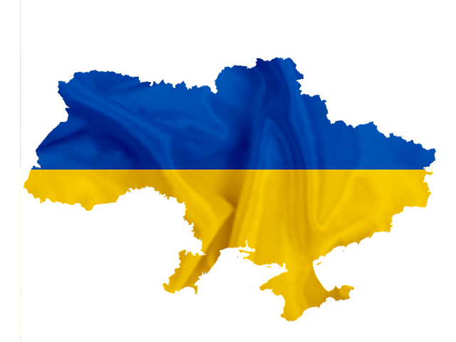
We warmly welcome our first member from Ukraine, Dr. Borys Olifirov, from the Bogomoletz Institute of Physiology of NAS of Ukraine who just joined Working Group 2, 5, 6, 7 and 8.
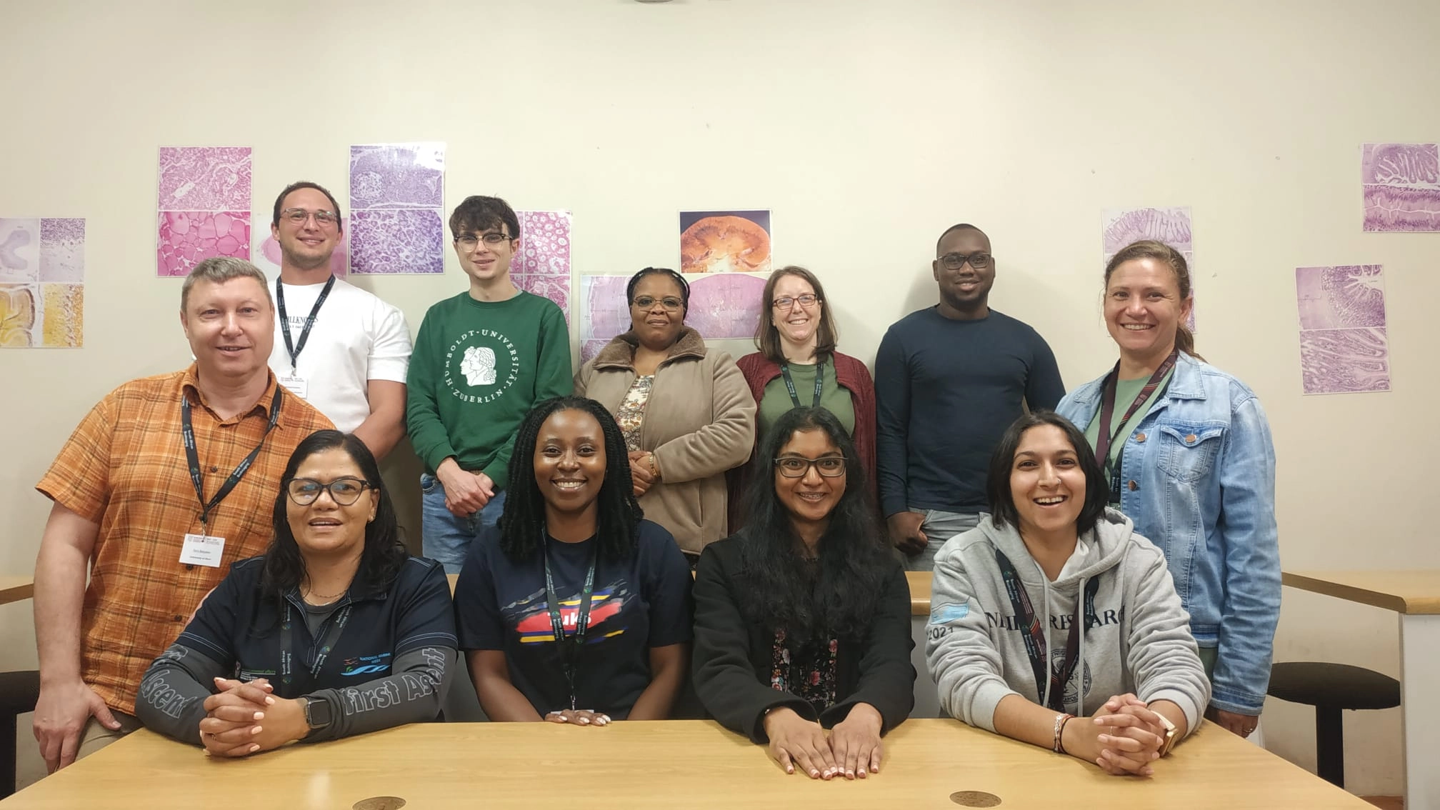
In April 2025, Yury Belyaev from the Microscopy Imaging Center (MIC) at the University of Bern, and a member of QUAREP-LiMi Working Group 5 (WG5), visited the Central Analytical Facilities (CAF) Microscopy Unit at Stellenbosch University, South Africa, as part of the Global BioImaging (GBI) International Job Shadowing Program During his visit, Yury Belyaev collaborated […]
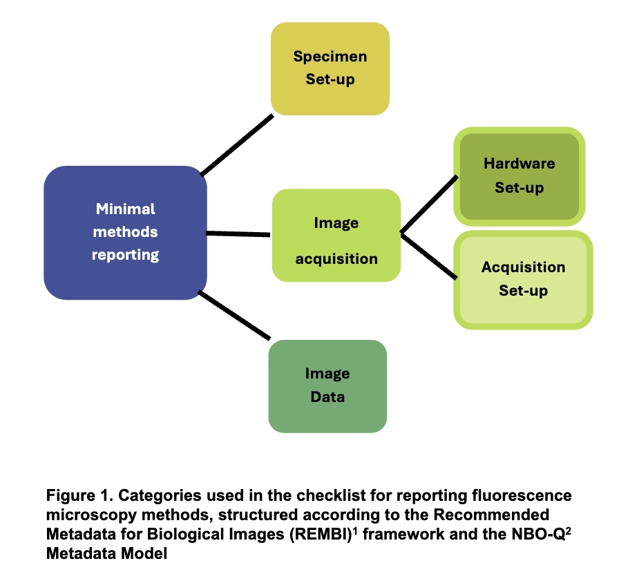
WG11, as part of its mission to enhance publication standards for reproducible image-based science and in response to an inquiry from Nature Methods, has defined the “Bare Minimum Requirements for Microscopy Methods Reporting“. This checklist, built upon the REMBI framework and the NBO-Q metadata model, is designed to guide researchers, scientific journals, and editors in […]